You can download this poster/image in high quality. You can then print it at your local printing shop and hang it on the wall.
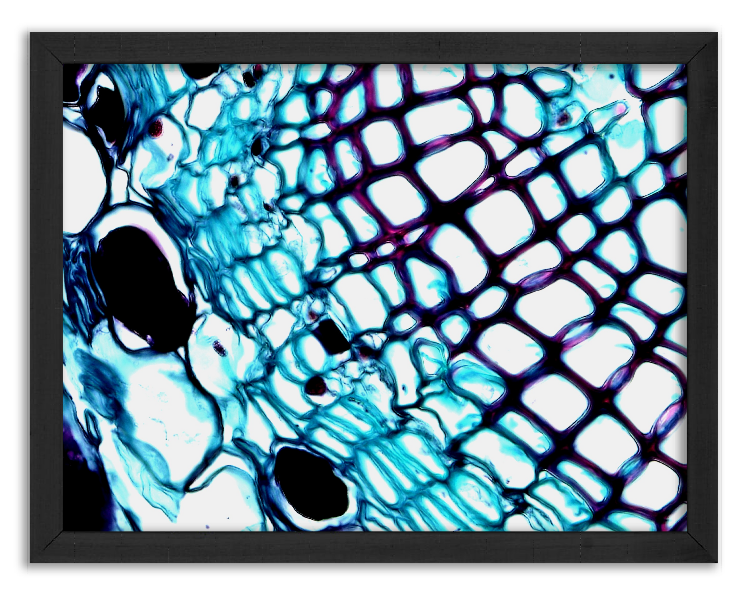
The illustration shows a longitudinal section of a pine (Pinus L.) root under a microscope at a magnification of x400. This root is composed of multiple tissue layers that serve to absorb water and mineral salts from the soil and transport them to other parts of the tree. Several characteristic structures can be distinguished in the pine root, including cork, cortex, phloem, and xylem layers. The cork layer protects the root from water loss and external factors, such as pathogens and mechanical damage. The cortex layer stores reserve substances and conducts water and mineral salts. The phloem serves to transport water and organic substances up the tree, while the xylem provides mechanical support and stores reserve substances. The pine root is well adapted to live in harsh soil and climate conditions, making it one of the most important organs of this tree.
You can download this poster/image in high quality. You can then print it at your local printing shop and hang it on the wall.
Subscribe to the newsletter. You will be the first to know about newly added articles or images for free download!

We are passionate about the world seen under the microscope. We want to share our passion with others.